THE BEST SOLUTION FOR
EACH INDIVIDUAL CASE
ANALYSES AND EVALUATIONS TO GET
A PREVIEW OF THE RESULTS OF THE SURGERY

Home/Image analysis/What it is
The examination of the person’s image entails the study of the individual’s general characteristics (age, gender, profession, sportive activities and all the general aspects of the individual’s personality) together with the physical features of the face.
Along with these elements, aspects related to nasal functions are not overlooked since they are very important for correct breathing.
The study of these elements will evidence the useful factors to take into consideration upon intervening on the single parts of the face, without foregoing the concepts and criteria of esthetic beauty. The study of the image uses photographic material and computerized simulations aimed at showing the patient the images that reproduce the final results of the operation. Obviously, the evaluation of the solution to be taken will be agreed on between the surgeon and patient.
The next step is, therefore, the study of the right-side and left-side profile (differs from the prior study), so as to furnish the patient with more than one solution. The patient will assess the differences between the various solutions proposed and will choose that which he/she prefers most. In some cases, 5/6 simulations are done of the final result, to be absolutely sure that the final proposal of the image fully satisfies the surgeon (in the sense of the technically achievable results) and obviously, the patient.
Once the correct solution is found, this is overlapped with the original image so that the patient can have a concrete vision of the extent of the corrections.
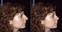
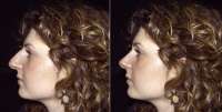
The two photographs laid side by side above, first show a curved nose profile compared to the simulation of the result where a linear profile is proposed. In this case, the study was done on the patient’s right-side profile.
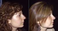
The two images below instead shows the left-side profile, proposed alongside a photo of the patient bearing the simulation of postoperative results.
The simulation of the two profiles differ, in order to give the patient two situations that respect the normal situation but at the same time offer different features and characteristics. In this case, the patient chose the right-side profile.
The two images below show two different situations: the first photo shows the overlapping of the real image with that of the simulation, allowing the patient to assess in a tangible manner the extent of the changes. The next photo shows the patient after the rhinoplasty treatment. The postoperative result demonstrates how true and reliable the study of the image was, and how the prospective solution substantially coincided with the results achieved.


